
Decoding the Image Part II: Understanding your knee imaging
Why the knee? Well, time and again, patients confide in me that they find themselves nodding along as their doctor points to MRI images, while secretly thinking, "What am I actually looking at here?" Sound familiar? We've all been there! That's exactly why we created this newsletter – to bridge the gap between medical imaging and understanding.
In this issue, we'll walk you through everything you need to know about knee X-Rays and MRIs in clear, simple terms. From ligaments to menisci, cartilage to bone, we'll help you understand what those mysterious gray and white images really mean for your health. Let's demystify your knee imaging together!
Understanding X-Rays: The Basics
X-rays show the bones of your knee joint. Think of them as shadows – dense structures like bones appear white, while soft tissues appear in shades of gray, and air appears black. They're particularly useful for seeing alignment, arthritis, and fractures.

Pictured left: A normal X-Ray of the knee
Key Landmarks on Knee X-Rays
The Three Main Bones
Femur (thighbone) – the large bone at the top
Tibia (shinbone) – the large bone at the bottom
Patella (kneecap) – the circular bone in front
Joint Spaces
Medial (inner) compartment
Lateral (outer) compartment
Patellofemoral (kneecap) compartment
Growth Plates (in children)
Appear as dark lines through the bones
Typically closes at the time of puberty
Different X-Ray Views
Front View (AP)
Shows overall alignment
Best for seeing joint spaces
Shows arthritis severity
Side View (Lateral)
Shows kneecap position
ACL injuries may show subtle signs
Shows fluid in joint
Sunrise/Merchant View
Shows kneecap tracking
Reveals patellofemoral arthritis
What we are looking for on a Knee X-Ray
Narrowing of the joint space on one or both sides
Irregular (or jagged) surfaces of the joint
Obvious breaks or fractures
Normal alignment of bones and positioning of the patella
Take a look at the X-Ray picture below and see if you can tell what is wrong. Hover over the picture with your cursor to reveal the answer.

The patella is dislocated laterally, it should be in the center of the knee
Look at the X-Ray below and take a guess on what you are looking at.
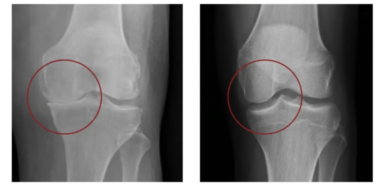
Notice the decrease in medial (inside) joint space in the picture on the left. This indicates an arthritic knee. The right is a normal X-Ray.
Understanding your MRI: A Detailed View
MRI’s are challenging to read, even for the most trained eyes. You really need to know your anatomy to decipher what you are looking at. There is a website that helps label everything as you scroll listed below, and I encourage everyone to take a look. Remember, MRIs shows soft tissues that X-rays can't capture. Each image is like a slice through your knee from different angles.
Here is a lateral view of the knee, meaning we are looking at the knee from the side with the slice from almost the exact middle of the knee.

Here is an axial view of the knee, meaning we are looking directly above your knee, right in the middle of the joint.

How to Read MRI Colors
There are 2 different types of MRI’s; T2 where the bone & fluid are light colored, and T1 where the bone and fluid is dark).
For example: In a T2- weighted MRI:
Black = Air and dense bone
White = Fluid, swelling, and some tendons
Gray = Most muscles and normal tissues
Dark Gray = Tendons and ligaments
Key Structures to Identify on MRI
Menisci (In the joint space; absorbs impact and stabilizes the knee)
Appear as dark triangular structures
Located between femur and tibia
One on inner (medial) and outer (lateral) side
Should be uniformly dark without breaks
Ligaments
ACL (Anterior Cruciate Ligament)
Appears as a dark diagonal band in the center
Should be continuous without gaps
PCL (Posterior Cruciate Ligament)
Appears as a dark "C" shape behind the ACL
MCL (Medial Collateral Ligament)
Dark band on inner side of knee
LCL (Lateral Collateral Ligament)
Dark band on outer side of knee
Cartilage
Appears as a bright white layer covering bone ends
Should be smooth and even
Visible on both femur and tibia
Patella (Kneecap) Tracking
Position relative to groove in femur
Thickness and condition of cartilage underneath
Common Abnormal Findings
Meniscus Tears
White line going through normally dark meniscus
Irregular shape or fragment
May see "parrot beak" appearance
ACL Tears
Gap in the normal dark band
Wavy appearance
Fluid (white) where ligament should be
Arthritis
Narrowed joint spaces on X-ray
Bone spurs (osteophytes)
Irregular cartilage surface on MRI
Patellofemoral Problems
Abnormal tracking of kneecap
Cartilage wear under kneecap
Tilting of the patella
Tips for Viewing Your Images
Understanding Orientation
Remember: Left side of image is usually your right side
Top of image is usually the front of your knee
Dark joint spaces should be even
Questions to Ask
"What is this dark/white area?"
"How does this compare to normal?"
"Can you show me where the problem is?"
Again, I urge you to take a look at this awesome website if you need help navigating your knee MRI.
Red Flags to Look For
On X-rays:
Uneven joint spaces
Bone spurs or loose bodies
Abnormal alignment
Fracture lines
On MRI:
Bright white areas in normally dark structures
Breaks in continuous structures
Large amounts of fluid
Bone marrow changes (signal changes within the bone)
Special Considerations
For Athletes
Focus on meniscus and ligament integrity
Look for bone stress reactions
Cartilage damage patterns
For Older Adults
Arthritis progression
Bone quality
Alignment issues
Remember: While this guide helps you understand your knee imaging, always rely on your healthcare provider's interpretation for medical decisions. Different imaging machines and settings can produce slightly different appearances, so some variation is normal.
Examples of Abnormal Scans
Now, you play the role of the radiologist. After reading and learning everything above, give it your best shot! The answer will reveal when you hover over the image.

MRI of an ACL tear. Notice the ligament is not attached on the bottom bone (tibia)

MRI of a meniscal tear. Notice the irregularity in the lateral (outside) meniscus.

The change in color consistency within the femur indicates edema, or in this case a bone bruise.